With this procedure, it is possible to identify and remove the entire tumour, while preserving healthy skin around the lesion. This technique consists of removing the cancer from the skin, layer by layer and examining each one under the microscope, until a free margin is obtained, that is, until the tumour has been completely removed (the level of precision and accuracy can reach 98%). This precision is possible since during the surgical procedure practically 100% of the margins are analysed by microscope. Once the free margin is reached, the wound (resulting from the extraction of the tumour) is reconstructed.
The main advantage of Mohs Micrographic Surgery when compared to conventional surgery is related to the microscopic control of the margins of the tumour during surgery. Removal of the entire tumour without aggression or extraction of normal skin is ensured. In conventional surgery, the tumour is removed with safety margins and sent to pathological anatomy. The result is usually received within 1-2 weeks. The risk of some cancerous residue remaining may exist and is definitely higher in relation to Mohs surgery, as removal of the tumour is carried out by what can be visualized by the dermatologist.
This issue of “extending the safety margin of the extraction” has always been much discussed in dermatological surgery; exiguous margins tend to leave remnants of the tumour, but facilitate reconstruction, while enlarged margins tend to completely remove tumours, but they can produce functional or even aesthetic sequelae, in addition enlarged surgical margins do not always guarantee total tumour removal. In short, the concept of “safety margin” is based on the supposed prediction of subclinical tumour growth, which, in reality, cannot be anticipated by a basic examination only.
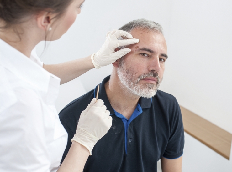
In order to perform Mohs micrographic surgery, it is necessary that the specialist has a deep knowledge of skin histology – to permit a microscopic analysis of the skin during surgery, to ensure a complete removal of the cancer, even in areas that are not clinically visible. He must also have surgical knowledge and most importantly reconstruction techniques.
The name micrographic surgery refers to the precise mapping and orientation performed during Mohs surgery, which allows the tumour to be removed and, at the same time be examined (described above). The term Mohs refers to the name of the creator of the technique, Frederic E. Mohs, who began the procedure in the 1930s. However, with the technological development in medicine, this technique has undergone a huge transformation, especially with the use of the cryostat, a device that allows skin slicing and freezing, so that the tumour can be examined during surgery.
It can also be indicated for basal cell carcinomas considered to have a low risk of recurrence, when the objective is to preserve healthy skin. Whether to reduce the size of the scar, or for areas where there is no excess skin to perform the reconstruction, as is the case, for example, in the auricular regions (ears), eyelids and glans penis.
Mohs surgery is indicated for:
- Basal cell carcinoma with an increased risk of recurrence;
- Squamous cell carcinoma (or squamous cell);
- Dermatofibrossarcoma protuberans (DFSP);
- And some other rare skin tumours.
Dr. Tiago Mestre is one of the few Portuguese Dermatologists who performs Mohs surgery, whose skills he acquired at Newcastle Upon Tyne Hospitals (United Kingdom). Dr. Tiago Mestre is a member of the American College and the European Mohs Surgery Society.


